Anatomi Platysma Pada Tubuh Manusia - SIPAT. Google Scholar Doty RW Bosma JF 1956 An electromyographic analysis of reflex deglutition.
![]()
Platysma Muscle Attachments Innervation Function Kenhub
Kayla LaFrance on Twitter.
Platysma-Ursprung und Insertion. Nevertheless the exact number is difficult to define. CourseAnatomy and Histology ANAT2008. The margins of the platysma can be held apart with.
Das Platysma gehört zur oberflächlichen Schicht der ventralen HalsmuskulaturAls dünne Muskelplatte bedeckt es nahezu die gesamte anteriore Fläche des Halses und wird funktionell zur mimischen Muskulatur gerechnet. Lower m argin of. The rectus capitis posterior major or rectus capitis posticus major both being Latin for larger posterior straight muscle of the head arises by a pointed tendon from the spinous process of the axis and becoming broader as it ascends is inserted into the lateral part of the inferior nuchal line of the occipital bone and the surface of the bone immediately below the line.
They are divided into three regional groups from superior to inferior. Platysma Draws outer part of lower lip. Occipitalis Muscle is an important muscle responsible for facial movements.
The occipital section or the belly of the epicranius muscle helps an individual to extend the scalp such that the eyebrows may come up. The fibres passed craniolaterally from this origin in the midline. Das Platysma weist einen großen Variationsreichtum auf.
Das Platysma das SMAS und die Faszia. This is a table of skeletal muscles of the human anatomy. The platysma belongs as all other facial muscles to the group.
Start studying Thoracic Muscles origins and insertions. Mit Ausnahme des M. The spinalis muscles are the most medial erector spinae muscles.
The muscle helps move the scalp and wrinkle the forehead as well as raise the eyebrows. Zygomatici major et minor haben ihren Ursprung am Knochen dh. Even though the occipitalis muscle is located at the back base of the skull many of the functions and actions of this muscle impact areas in the front of.
Sie inserieren mit kurzen zumeist elastischen Sehnen in der Subkutis oder im Korium der Haut. Variations are those of origin and insertion eg from the external occipital protuberance and insertion into the parotid fascia and the angle of the mouth interdigitaying with platysma. Muscle aro und.
Separated from the platysma cranially caudally and medially and the platysma is also separated following the direction of its fibres. Spinalis capitis muscle originates from the spinous processes of C7-T1 vertebrae and inserts into the midline of the occipital bone. Ame of Muscle Actions Origin Insertion Head Face Neck.
The trapezius muscle has several origin points along the midline of the posterior neck and back. Almost every muscle constitutes one part of a pair of identical bilateral muscles found on both sides resulting in approximately 320 pairs of muscles as presented in this article. Es kann als dünne blasse Muskelplatte mit zahlreichen bindegewebigen Unterbrechungen.
Platysmaplasty is a rejuvenation procedure performed to increase the definition of the neck from the angle of the jaw to the chin thereby restoring a youthful and aesthetic contour to the face. Elliott DM Robinson PS Gimbel JA Sarver JJ et al. The biceps brachii muscle biceps is a large thick muscle of the arm consisting of two heads.
The temporalis muscle runs superficially from the temporal bone to the coronoid process of mandible. Most of the fibres arose from the midline between the occipital bone and the first thoracic vertebra. Its unique morphology structure and variations have drawn genuine interests in this muscle from anatomists scientists and physicians for a long time and the variations of the digastric muscle have been documented since the 18th century.
Die meisten mimischen Muskeln haben ihren Ursprung am Schädel oder an den Aponeurosen und Bindegewebssepten. Learn vocabulary terms and more with flashcards games and other study tools. The superior fibers attach on the medial third of the superior nuchal line and the external occipital protuberance of the occipital boneThese fibers have a descending course towards their insertion point hence why this part of the trapezius is referred to as the descending part.
Temporalis muscle Musculus temporalis The temporalis muscle is a thin fan-shaped muscle situated within the temporal fossa of the skullAlong with the medial pterygoid lateral pterygoid and masseter muscles it belongs to the group masticatory muscles. Insertion into the angle of the mouth allows it to act as a risorius muscle. Pectoralis major muscle Musculus pectoralis major The pectoralis major is a paired superficial muscle located on the anterior surface of the thoracic cageIf youre a gym lover youll hear these muscles also being referred to as the pecs musclesThe pectoralis major has a broad origin based on which it is divided into three parts.
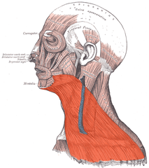
It completely covers both front sides of the neck. Anatomy origin insertion function Kenhub. über ursprung und insertion der skeletmuskein.
Sie befinden sich in ihren. NerdArmy DYK that the strongest muscle in the human body is the masseter jaw muscle Masseter Muscle. Buccinator weisen die mimischen Muskeln keine Faszienbe-deckung also kein Perimysium auf.
The long biceps tendon makes a sharp turn at the humeral head and continues its course in the bicipital groove intertubercular sulcus. The platysma is a superficially proceeding muscle and unlike other muscles directly connected to the skin. There are around 650 skeletal muscles within the typical human body.
Insertion and boundary of the TNAFs and the effect of. Inferiorly and posteriorly pouting. Effect of altered matrix proteins on quasilinear viscoelastic properties in transgenic mouse tail tendons.
Mandible skin and. Epicranius fr ontalis Closes eye F rontal and maxillary bones ligaments aro und orbit. Spinalis colli muscle originates from the same points as spinalis capitis but also from the nuchal ligament.
Dolgo-Saburoff B 192930 Über Ursprung und Insertion der Skelettmuskeln. The platysma was found to be a large thin quadrangular muscle that covered the sides of the neck and formed a thin layer of fibres closely related to the skin Fig. CAS PubMed Google Scholar.
Cranial und caudal anchoring elements in form of spikes for a. Due to the direct insertion to the skin it can change the facial expression along with other facial muscles. Clavicular part sternocostal part and abdominal part.
The digastric muscle consists of the anterior belly and the posterior belly connecting the mandible hyoid bone and temporal bone. Originates at the supraglenoid tubercle above the glenoid cavity of the scapulaIt lies within the intracapsular space but it still remains extrasynovial.
File Platysma Png Wikimedia Commons
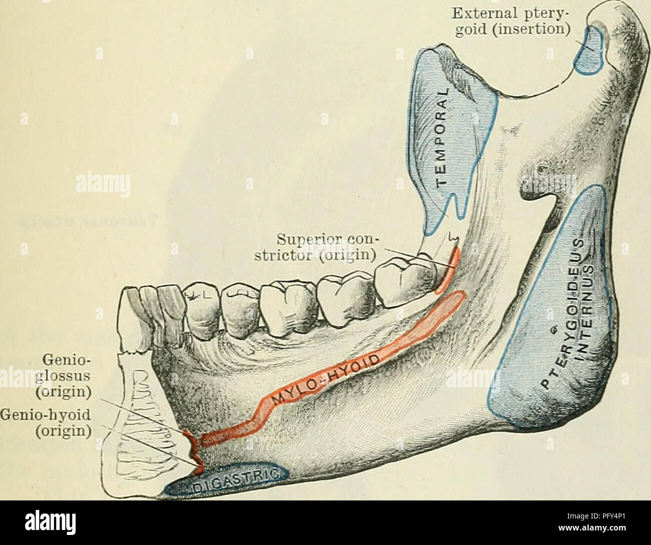
Cunninghams Lehrbuch Der Anatomie Anatomie M Triangularis Ursprung M Quadratus Labii Inferioris Origin M Mentalis Ursprung Platysma Av E Insertion Abb 403 Muscle Attachments Zur Lateralen Aspekt Des Unterkiefers Nach Unten